Ultrasound imaging uses high-frequency sound waves to produce real-time images of internal body structures — without ionizing radiation, contrast agents, or the constraints that make MRI inaccessible for some patients. It is one of the most versatile, accessible, and safe imaging modalities in clinical medicine. Point-of-care ultrasound (POCUS) — performed directly by the clinician at the bedside — is increasingly available even in primary care settings. This guide explains the broad range of clinical conditions diagnosed and monitored through ultrasound.
Obstetric Ultrasound
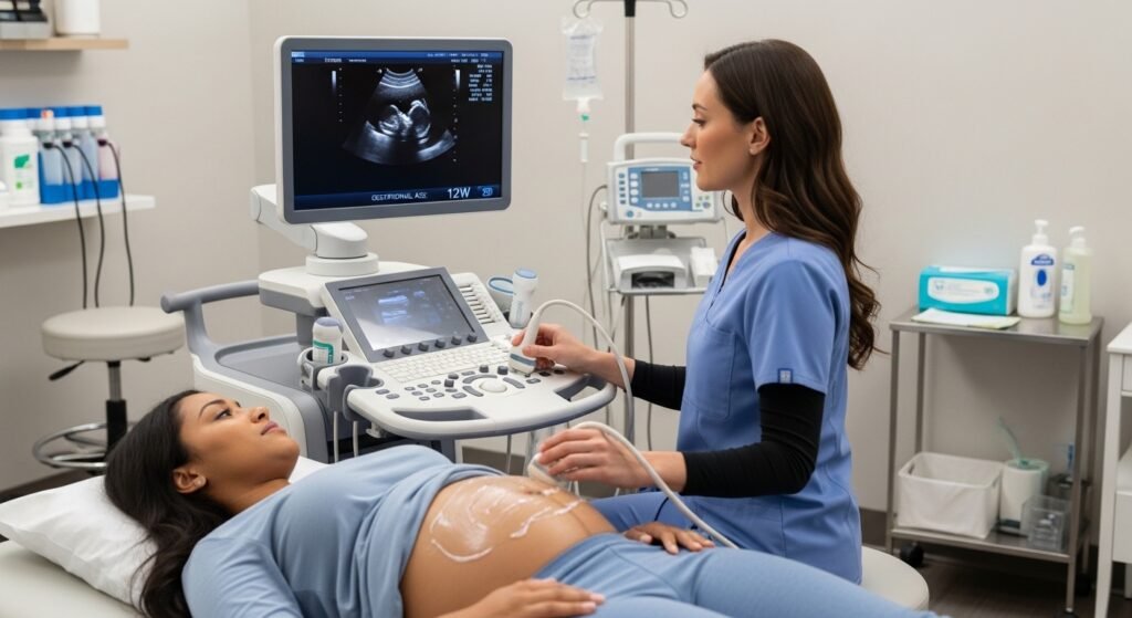
Pregnancy ultrasound is perhaps the most recognized application — confirming viable intrauterine pregnancy, measuring gestational age and fetal growth, assessing placental position, detecting structural fetal anomalies (targeted anatomy scan at 18–20 weeks), and evaluating amniotic fluid volume and fetal well-being. Obstetric ultrasound is a fundamental component of prenatal care.
Abdominal and Pelvic Ultrasound
Abdominal ultrasound evaluates liver (fatty liver, cirrhosis, masses), gallbladder (gallstones — the primary indication), bile ducts, kidneys (stones, hydronephrosis, cysts, masses), spleen, pancreas (masses, ductal dilation), and abdominal aorta (aneurysm screening). Pelvic ultrasound evaluates the uterus, ovaries, and bladder for fibroids, ovarian cysts, and other pelvic pathology.
Musculoskeletal Ultrasound
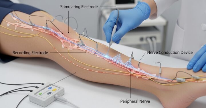
Ultrasound is increasingly used for dynamic assessment of tendons, muscles, and joint spaces — visualizing rotator cuff tears, tennis elbow tendinopathy, joint effusions, and Baker’s cysts. Ultrasound-guided joint injections and aspirations provide superior accuracy compared to landmark-based technique.
Vascular Ultrasound
Doppler ultrasound measures blood flow through arteries and veins — diagnosing DVT in leg veins, carotid artery stenosis, peripheral arterial disease, and heart valve function. Duplex ultrasound combines structural imaging with blood flow assessment.
Conclusion
Ultrasound is a cornerstone diagnostic tool across medicine — safe, portable, real-time, and radiation-free. Its expanding use at the point of care by trained clinicians is bringing diagnostic imaging directly to the patient’s bedside, reducing time to diagnosis and improving clinical efficiency. When your clinic orders an ultrasound, it is choosing an appropriate imaging tool for a specific clinical question with an excellent safety profile.
FAQs – Ultrasound
Q1. Is ultrasound safe during pregnancy?
A: Yes. Diagnostic ultrasound has been used in obstetric care for over 50 years with no demonstrated adverse effects to mother or fetus at standard diagnostic intensities. It is the preferred imaging modality throughout pregnancy.
Q2. Why does ultrasound require gel?
A: The gel eliminates air between the transducer and skin — air reflects sound waves, preventing them from entering the body. The gel conducts sound waves effectively, enabling clear image transmission.
Q3. Can ultrasound detect cancer?
A: Ultrasound detects many cancers — thyroid, breast, ovarian, liver, kidney. It is particularly useful for characterizing masses found on other imaging and guiding biopsy. For some cancer types (e.g., solid versus cystic breast masses), it provides diagnostic information complementary to mammography.
Q4. Is an endoscopic ultrasound the same as a regular ultrasound?
A: No. Endoscopic ultrasound (EUS) uses a flexible scope with an ultrasound probe at the tip, inserted through the mouth or rectum, providing close-range imaging of structures near the GI tract — pancreas, bile ducts, lymph nodes, rectal wall — with superior detail compared to external ultrasound.
Q5. Does ultrasound show kidney stones?
A: Ultrasound detects kidney stones within the kidney well but misses smaller stones in the ureter. CT without contrast is the most sensitive imaging for ureterolithiasis. Ultrasound is a reasonable first-line study for suspected kidney stones, particularly to minimize radiation exposure in appropriate patients.