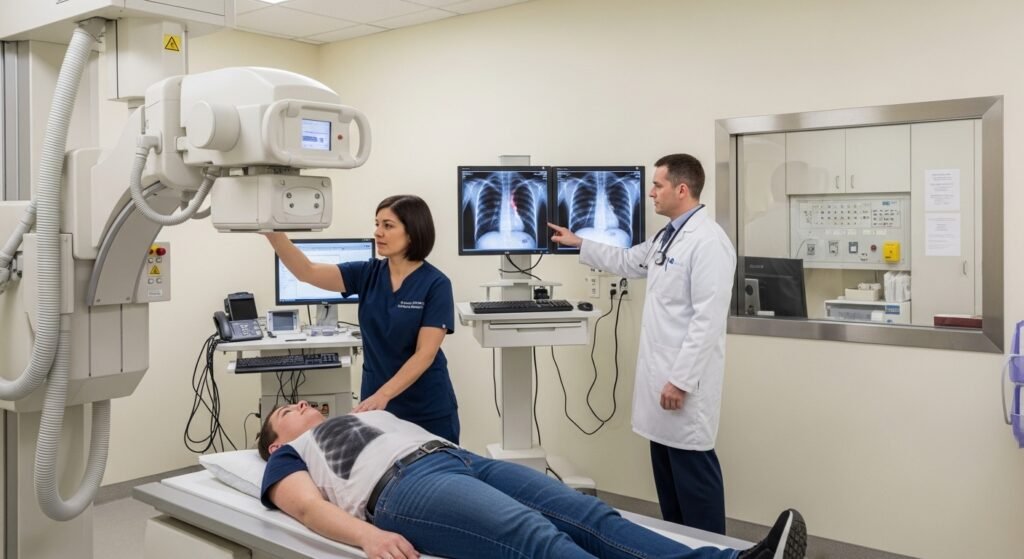
X-rays (radiographs) are the most widely used diagnostic imaging modality in medical clinics — fast, inexpensive, widely available, and providing essential diagnostic information for a broad range of conditions. Despite being over 125 years old, X-ray technology remains fundamental to everyday clinical medicine. Understanding what X-rays show and when they are most appropriate helps you appreciate the clinical reasoning behind a clinic’s imaging decisions. This guide explains clinical X-ray use.
How X-Rays Work
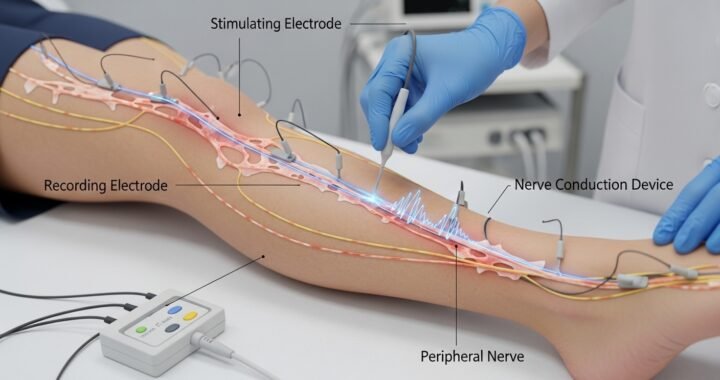
X-rays are electromagnetic radiation that passes through body tissues to varying degrees depending on tissue density. Dense structures (bone) absorb X-rays, appearing white. Air-filled structures (lungs) allow passage, appearing black. Soft tissues appear in intermediate shades of gray. This differential absorption creates the image that radiologists and clinicians interpret for anatomical and pathological information.
Common Clinical Uses
Chest X-Ray
The chest X-ray is one of the most frequently ordered studies — assessing lung fields (pneumonia, pleural effusion, pneumothorax, masses), heart size (cardiomegaly), mediastinal anatomy, and bony structures of the chest wall. It is a rapid, low-dose screening tool for respiratory and cardiac conditions.
Skeletal X-Rays
Fractures, dislocations, degenerative joint disease, bone tumors, and inflammatory arthritis changes are all evaluated through skeletal radiographs. X-rays of specific joints — knee, hip, shoulder, hand — provide important structural information before and after orthopedic intervention.
Abdominal X-Rays
Plain abdominal X-rays assess bowel gas patterns (obstruction, ileus), radio-opaque foreign bodies, calcifications (gallstones, kidney stones visible on plain film), and free air under the diaphragm (indicating perforation). They have largely been supplemented by CT for most abdominal indications but remain useful for specific clinical questions.
Radiation Considerations
The radiation dose from a single X-ray is very low — a chest X-ray delivers approximately 0.1 mSv (equivalent to about 10 days of background radiation). The risks are minimal for individual studies, but cumulative radiation exposure over a lifetime, and radiation sensitivity in children and pregnant women, appropriately influence imaging decisions.
Conclusion
X-rays remain a fundamental, efficient, and low-cost first-line imaging tool for a wide range of clinical conditions. They provide valuable diagnostic information rapidly and with a very low radiation dose — making them an appropriate first-line study for fracture evaluation, chest symptoms, and many other clinical indications where their diagnostic value clearly outweighs the minimal radiation exposure.
FAQs – X-Rays at Clinics
Q1. Are X-rays safe during pregnancy?
A: While no radiation is ideal during pregnancy, the radiation from a single X-ray is very low. If an X-ray is clinically necessary, it can generally be performed safely, particularly after the first trimester, with abdominal shielding. Always inform your clinician and the radiology staff of your pregnancy.
Q2. Why do I have to wear a lead apron for some X-rays?
A: Lead shields protect radiation-sensitive areas (gonads, thyroid, developing embryo) not included in the area being imaged. The shield reduces radiation to areas where the risk of harm is greatest while allowing diagnostic quality in the area of interest.
Q3. Do dental X-rays count toward radiation exposure?
A: Yes, though their dose is tiny (0.005 mSv per bitewing film — equivalent to less than a day’s background radiation). Modern digital dental X-rays use even less radiation. Their cumulative contribution to lifetime radiation dose is negligible compared to CT or nuclear medicine studies.
Q4. What is a “portable X-ray”?
A: A mobile X-ray machine that brings imaging to patients who cannot be transported to the radiology department — bedbound inpatients, residents in long-term care facilities, and homebound patients. Portable X-ray image quality is slightly lower than department equipment due to technical constraints.
Q5. How quickly are X-ray results available at a clinic?
A: In clinics with on-site radiology services, preliminary results may be available within minutes and formal radiologist interpretation within hours. Clinics ordering from external radiology centers typically receive formal reports within 24–48 hours, with critical findings communicated immediately by phone.