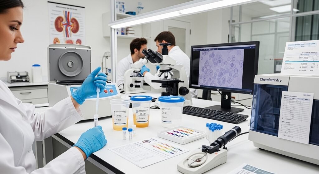
Urine tests — collectively called urinalysis — are among the most informative, least invasive, and least expensive diagnostic tests available at medical clinics. A routine urinalysis can detect kidney disease, urinary tract infection, diabetes, liver disease, and even signs of certain cancers. Understanding what urine tests measure and how clinics interpret the results makes you a more informed participant in your own care. This guide explains urinalysis and related urine testing at the clinical level.
Components of a Routine Urinalysis
Dipstick Analysis
A chemically treated dipstick is dipped in urine and produces color changes indicating: glucose (elevated in diabetes), protein (indicates kidney damage or glomerular disease when persistent), leukocyte esterase and nitrites (white cells and bacteria — indicating UTI), blood (hematuria — may indicate infection, kidney stones, or less commonly malignancy), ketones (present during fasting, diabetic ketoacidosis, or low-carbohydrate diets), bilirubin and urobilinogen (liver function markers), and specific gravity (measures urine concentration reflecting hydration).
Microscopic Examination
When the dipstick suggests abnormality, a microscopic urine examination identifies specific cell types (red blood cells, white blood cells, epithelial cells), casts (protein or cell aggregates indicating specific kidney disorders), and crystals (found in kidney stone formers). This microscopic assessment provides significantly more diagnostic information than the dipstick alone.
Urine Culture
When UTI is suspected, a urine culture identifies the specific causative organism and its antibiotic sensitivities — guiding targeted antibiotic selection. Culture results typically take 24–72 hours. Mid-stream clean-catch technique is essential for accurate results — contamination with vaginal or perineal bacteria produces false positives that can lead to unnecessary antibiotic treatment.
24-Hour Urine Collection
For certain assessments — kidney stone risk profiling, protein quantification in kidney disease, cortisol measurement for Cushing’s syndrome — a 24-hour urine collection (collecting all urine over 24 hours) provides more accurate quantitative data than a single specimen.
Conclusion
Urine tests provide a remarkable window into kidney, metabolic, and urological health — and they are simple to collect and perform. Understanding what your urinalysis results mean helps you appreciate the value of this simple test and identify patterns that warrant attention. When your clinic orders a urine test, provide a clean mid-stream specimen per the instructions provided — sample quality significantly affects result accuracy.
FAQs – Urine Tests
Q1. What does protein in urine mean?
A: Small amounts of protein in urine (microalbuminuria) are an early sign of kidney damage, particularly in diabetic and hypertensive patients. Larger amounts (overt proteinuria) indicate more significant glomerular damage. Persistent protein in urine warrants further kidney function evaluation.
Q2. What causes blood in urine?
A: Hematuria (blood in urine) has many causes ranging from benign (vigorous exercise, menstrual contamination, urinary tract infection) to serious (kidney stones, bladder cancer, kidney disease). Any unexplained blood in urine in an adult warrants clinic evaluation — particularly in patients over 50 or with risk factors for bladder cancer.
Q3. Does drinking water before a urine test affect results?
A: Adequate hydration produces a properly concentrated sample. Excessive hydration dilutes urine, potentially causing false-negative results for glucose, protein, and other analytes. Drink normally before routine urinalysis unless given specific instructions.
Q4. Can medications affect urine test results?
A: Yes. Many medications alter urine color, pH, and chemical reactions on dipstick — nitrofurantoin (brown), rifampin (orange-red), metronidazole and some B vitamins (dark brown or green). Inform your clinic of all medications when providing urine samples.
Q5. How do I collect a clean mid-stream urine sample?
A: Clean the genital area with the provided wipes, begin urinating into the toilet, then collect the mid-stream portion in the specimen cup. This technique reduces contamination with perineal bacteria and skin cells, improving the accuracy of culture results.